Immunohistochemistry (IHC)

Every Triple A Polyclonal and PrecisA Monoclonal antibody is validated by immunohistochemistry (IHC) on formalin-fixed, paraffin-embedded (FFPE) human tissues in tissue microarray (TMA) format.
576 tissue cores per antibody
IHC is a qualitative method, so our experts review every tissue core manually. For each tissue, they record:
To further verify specificity, the validation performed for our antibodies is expanded with application-specific Enhanced Validation.
In IHC, two different Enhanced Validation methods are applied:
1. Orthogonal validation (verification done with a non-antibody-based method
2. Independent antibody validation (two antibodies targeting the same protein but binding to different parts of the protein thus verifying each other)
Currently, over 5,000 antibodies are validated using at least one of these two enhanced validation methods. Learn more about the methods below.
👀 Watch the animation about Enhanced Validation
1. Orthogonal validation compares antibody signal to RNA
Orthogonal validation in IHC involves confirming antibody staining using a non-antibody-based method, like RNA-Seq. In IHC, this means comparing the antibody signal to RNA data from the same samples.
For validation, two tissues are selected for each antibody: one with high RNA expression and one with low or no expression of the target protein. The RNA levels in the two tissues should differ by at least five times.
The validation results show both the IHC staining for these two samples and their corresponding RNA data.
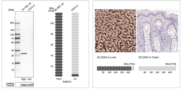
Example of orthogonal validation in IHC. IHC staining of liver and kidney tissues using the Anti-SLC2A2 polyclonal antibody (HPA028997). The corresponding RNA-Seq data (TPM values) for the same tissues are presentedat the bottom. Liver and colon samples were chosen because of their high and low SLC2A2 RNA expression, respectively.
2. Validation by independent antibody
This validation method involves confirming antibody specificity by comparing at least two antibodies that target the same protein but recognize different epitopes.
If the staining patterns from both antibodies are consistent across a range of relevant tissues, they validate each other. At Atlas Antibodies, 20-44 tissues are tested for each antibody, and four representative tissues are shown for each one.
The validation results include IHC staining images for both antibodies side by side.
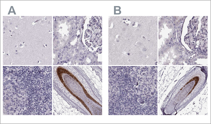
Example of validation by independent antibodies in IHC. The two Anti-TCHHL1 antibodies HPA063483 (left, A) and HPA042579 (right, B) target different regions of TCHHL1. The image shows IHC stainings of the two antibodies in the cerebral cortex, kidney, lymph node, and skin tissues. The antibody's staining pattern across positive and negative tissues is comparable, so the two antibodies validate each other.
Get the best results with protocols optimized for our Triple A Polyclonals and PrecisA Monoclonals. Choose the standard protocol for manual staining, or the Ventana Discovery XT protocol for automated staining. Both include practical advice on IHC validation and troubleshooting.
Discount code: "ATLAS20YEARS"
Valid through 2026.
Use code at checkout on atlasantibodies.com