Molboolean™
Simultaneous In Situ Detection of Free & Interacting Proteins


MolBoolean™ detects and quantifies free proteins and protein complexes in fixed cells and tissue. PLA shows only interactions. MolBoolean shows protein A, protein B and complex AB in a single experiment.
Researchers using standard In situ Proximity Ligation Assay (isPLA) face a common limitation: isPLA detects only A-B protein complexes. It does not show how much of protein A or protein B is free. So when the interaction signal drops, you can’t tell whether the complex dissociated or the protein was simply downregulated.
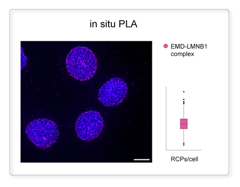
MolBoolean was designed to close that gap. Instead of reporting only where proteins interact, it detects the free, non-interacting proteins and the complex itself at the same time. You get the full picture in a single assay and can see what is really happening in the cell.
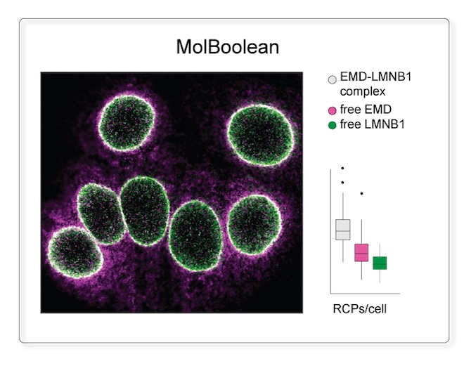
MolBoolean detects free protein A, free protein B and the A–B complex in the same cell, in one staining. You can measure the bound and unbound pool of each protein, and what fraction of it takes part in the interaction, at single-cell resolution.
Kit specification
MolBoolean Starter Kit – 1.6 mL (approx. 20 tissue assays or 40 cell assays)
MolBoolean Standard Kit – 4.8 mL (approx. 60 tissue assays or 120 cell assays)
The kit is customizable for most species upon request, providing flexibility for diverse research applications.
MolBoolean™ is not just an improvement.
It closes a fundamental measurement gap in spatial biology.
Discover why standard methods aren't enough. Principal Scientist Mikael Malmqvist, PhD, explains how MolBoolean provides clearer, more accurate data for protein-protein interactions.
Valid through 2026.
Use code at checkout on atlasantibodies.com
Human Kidney: ACE2 & TMPRSS2
MolBoolean™ analysis of the interaction (white) between ACE2 (magenta) and TMPRSS2 (green) in human kidney. Using anti-ACE2 (Cat. AMAb91259) and anti-TMPRSS2 (Cat. HPA035787) antibodies, the image displays the relative quantification of free versus interacting protein fractions. Results show 51% free ACE2, 17% free TMPRSS2, and 32% ACE2/TMPRSS2 complex, normalized to total target protein levels (total RCPs).
MCF7 Cells: E-Cadherin & β-Catenin
MolBoolean™ analysis of the interaction (white) between E-cadherin (cat. AMAb90862, magenta) and β-catenin (cat. HPA029159, green) in MCF7 cells, showing the relative quantification of free versus interacting protein fractions, indicated by the detection of rolling circle products (RCPs) in either one or two fluorescent channels: 32% free E-cadherin (magenta), 19% free β-catenin (green), 49% E-cadherin/β-catenin complex (white). Data is normalized to total target protein levels (total RCPs).
Human Colon: SATB2 & HDAC1
MolBoolean™ analysis of the interaction (white) between SATB2 (magenta) and HDAC1 (green) in human colon, using the monoclonal anti-SATB2 (Cat. AMAb90682) and the polyclonal anti-HDAC1 (Cat. HPA029693) antibodies.
Image shows the spatial location of free versus interacting protein fractions, indicated by the detection of rolling circle products (RCPs) in either one or two fluorescent channels.
MCF7 Cells: EMD & LMNB1
MolBoolean™ analysis of the interaction (white) between EMD (magenta) and LMNB1 (green) in MCF7 cells, using the monoclonal anti-EMD (Cat. AMAb90562) and the polyclonal anti- LMNB1 (Cat. HPA050524) antibodies.
Image shows the spatial location of free versus interacting protein fractions, indicated by the detection of rolling circle products (RCPs) in either one or two fluorescent channels.
How MolBoolean differs from PLA?
MolBoolean™ is a kit for in situ protein proximity analysis in tissue and cells developed by Atlas Antibodies. Unlike traditional methods, it provides spatial quantitative analysis of protein-protein interactions by the simultaneous detection of free and interacting proteins (~40 nm proximity).
The comparison is in situ PLA, and the difference is one sentence: PLA measures protein complexes; MolBoolean also measures the free proteins that are not in a complex — both, simultaneously, in the same cell. That’s not an incremental improvement; it’s a different data type.
What problem does it solve?
The classic misinterpretation in interaction studies: an increase in interaction signal can come from higher expression of the target proteins rather than an actual increase in interaction rate. PLA alone can’t distinguish these. MolBoolean can, because interaction data is normalized to total target protein levels.
The “Molecular Boolean" Logic
The MolBoolean™ assay applies a Boolean logic at a molecular level to distinguish between free and interacting proteins, mapping protein interactions with high spatial specificity.
The OR logic identifies either protein A or B independently, signaling that they are present but not necessarily interacting.
The AND logic is applied when proteins A and B are interacting; they are detected together as complex AB, indicating a combined "true" interaction.
What are the benefits of using MolBoolean™ compared to traditional methods for studying protein-protein interactions?
Comparative table that highlights the advantages of MolBoolean™ over in situ PLA and FRET for studying protein-protein dynamics:
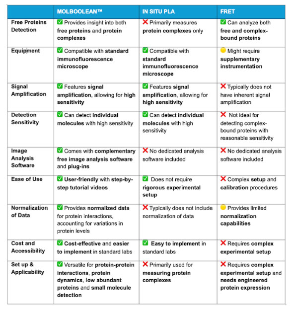
Why choose MolBoolean?
MolBoolean provides spatial quantitative analysis of protein-protein interactions by the simultaneous detection of free
and interacting proteins. MolBoolean™ offers significant benefits in fields requiring complex spatial interaction and quantification data such as:
Product Description
MolBoolean™ Mouse/Rabbit is a kit for in situ protein proximity analysis in tissue and cells. The MolBoolean™ assay utilizes a proprietary oligonucleotide setup that enables the simultaneous detection of both free and interacting (~40 nm proximity) fractions for two proteins of interest (protein A, protein B and interaction proteins AB). It uses two different immunofluorescent detection reporters with maximum emission wavelengths at 590 nm (ATTO565, TxRed filter or similar) and at 664 nm (ATTO647N, Y5 filter or similar), respectively.
MolBoolean™ Mouse/Rabbit can be performed with the user’s primary antibodies of choice (raised in mouse and rabbit). MolBoolean™ relies on anti-mouse and anti-rabbit secondary proximity probes and rolling circle amplification (RCA) as a mean to amplify signal.
A series of molecular steps, performed in the MolBoolean™ assay, incorporates information into the amplified products indicating antibody target engagement of individual versus interacting proteins. This information is converted to fluorescent signals by the binding of detection reporters, allowing visualization on a conventional fluorescent microscope. An image analysis software is provided to segment and differentiate fluorescent signals, enabling the relative quantification of free versus interacting fractions for the two analyzed protein targets in the sample.
MolBoolean Assay Steps and Workflow
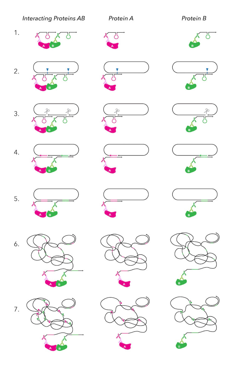
The MolBoolean™ assay follows a 2-days 7-steps workflow to determine if target proteins are interacting (AB) or present individually (A, B).
Day 1 (2 hours): blocking and primary antibodies incubation.
Day 2 (7-8 hours): probe binding, tagging, amplification, and detection.
(STEP 1) Proximity probes bind to primary antibodies attached to the proteins.
(STEP 2) Proximity probes link to the DNA circle.
(STEP 3) DNA nicking prepares the DNA for tagging.
(STEP 4) Tag oligos are added to mark the proteins.
(STEP 5) DNA ligation connects the DNA segments.
(STEP 6) Rolling circle amplification (RCA) boosts the signal for easier detection.
(STEP 7) Detection reveals both interacting and separate proteins.
What's Included in the MolBoolean Kit?
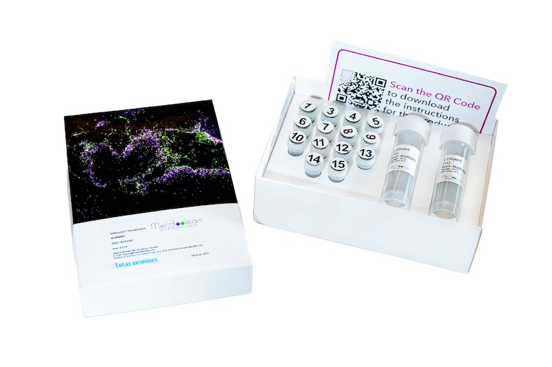
The MolBoolean™ kit includes 15 separate tubes: enzymes (nickase, ligase, polymerase), buffers, oligos for tagging and detection, secondary antibody probes and additional reagents such as blocker, and diluent.
1:Blocker (4x) - 2:Diluent (1x) - 3:Probe A (80x) - 4:Probe B (80x) - 5:Circle (40x) - 6:Additive (40x) - 7:Buffer A (10x) - 8:Nickase enzyme (80x) - 9:Buffer B (8x) - 10:Tag oligos (40x) - 11:Ligase enzyme (80x) - 12:Polymerase enzyme (40x) - 13:Buffer C (5x) - 14:Detection oligos (40x) - 15:Buffer D (10x).
The total volume of 4.8 ml, is sufficient for approx. 120 assays in cells (40 μl/assay) and 60 assays in tissue (80 μl/assay).
Can MolBoolean be used with any primary antibodies?
MolBoolean is a kit that can be used with the customer’s choice of primary antibodies (mouse /rabbit). MolBoolean is compatible with protocols already established in the lab for primary antibodies. This flexibility allows researchers to seamlessly integrate MolBoolean into their existing workflows without requiring the purchase of specialized antibodies, making it a convenient and adaptable tool for studying protein interactions and free protein levels.
Be aware that the specificity of the primary antibodies can affect the accuracy of MolBoolean results. It is important to validate the primary antibodies to ensure that they specifically target the proteins of interest before applying the MolBoolean technology. We recommend the use of Enhanced validated primary antibodies.
📖 Peer Reviewed Publications
Rivas-Santisteban R, et al, (2024) GPR88 impairs the signaling of kappa opioid receptors in a heterologous system and in primary striatal neurons. Neuropharmacology. 2024 Nov 27:110242. doi: 10.1016/j.neuropharm.2024.110242. Epub ahead of print. PMID: 39613254.
Kotliar IB, et al, (2024) Multiplexed mapping of the interactome of GPCRs with receptor activity-modifying proteins. Sci Adv. 2024 Aug 2;10(31):eado9959. Epub 2024 Jul 31. PMID: 39083597; PMCID: PMC11290489.
Malmqvist M, et al, (2024) Quantifying dopamine D2 and adenosine A2A receptor interactions in rat brain using MolBoolean™ technology. #8217 Society for Neuroscience, SfN, (2024).
Raykova D, et al, (2023) A method for Boolean analysis of protein interactions at a molecular level. Nat Commun. 2023 Sep 6;14(1):5450. doi: 10.1038/s41467-023-41325-3. Erratum for: Nat Commun. 2022 Aug 13;13(1):4755. doi: 10.1038/s41467-022-32395-w. PMID: 37673885; PMCID: PMC10482831.
Rivas-Santisteban R, et al, (2023) Boolean analysis shows a high proportion of dopamine D2 receptors interacting with adenosine A2A receptors in striatal medium spiny neurons of mouse and non-human primate models of Parkinson’s disease. Neurobiol Dis. 2023 Nov;188:106341.doi: 10.1016/j.nbd.2023.106341. Epub 2023 Oct 31. PMID: 37918757.
Gao X, et al, (2025) Molecular Boolean analyses of chemokine (C-C motif) receptor 1, α1B-adrenoceptor and arginine vasopressin receptor 1A heteromers. Biochem Biophys Rep. 2025 Dec 5;45:102404. doi: 10.1016/j.bbrep.2025.102404. PMID: 41867271; PMCID: PMC13005234.