
Every tissue section holds far more biological data than conventional staining methods can extract. Low-abundance proteins frequently get lost in background noise, and clashing same-species antibodies force researchers into grueling, multi-day cycles of sequential staining and harsh chemical stripping.
For researchers in oncology, neuroscience, and spatial biology where understanding the exact spatial relationships between multiple biomarkers is critical, these technical limits present a massive bottleneck.
Tyramide Signal Amplification (TSA) is the enzyme-mediated chemistry that eliminates these boundaries. It is also the core engine driving AtlasPlex™, a ready-to-use multiplex IHC/IF kit.
What is Tyramide Signal Amplification and How Does It Work?
In a conventional immunohistochemistry (IHC) or immunofluorescence (IF) stain, one primary antibody carries a fixed number of reporter molecules, making the visible signal directly proportional to antigen abundance. TSA breaks this one-to-one ceiling through enzymatic amplification.
The TSA Chemical Mechanism:
- Enzyme Binding: A primary antibody is targeted and linked to Horseradish Peroxidase (HRP).
- Substrate Activation: When a fluorophore-labeled tyramide substrate is introduced, HRP converts it into a highly reactive, short-lived intermediate.
- Covalent Anchoring: This active intermediate rapidly forms covalent bonds with tyrosine residues on and immediately adjacent to the target antigen site.
Because a single HRP enzyme deposits hundreds of tyramide-fluorophore molecules precisely at the target site, the result is a dramatically amplified, chemically permanent signal anchored directly to the tissue.
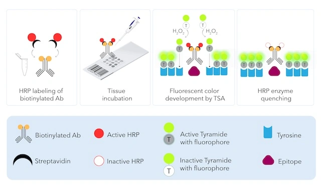
Fig.1 Enhanced Sensitivity for Low-Abundance Targets Tyramide signal amplification (TSA) works by tethering an enzyme to a specific antibody, then using that enzyme to deposit many dye molecules right at the target site. A biotinylated primary antibody is first linked to streptavidin-conjugated horseradish peroxidase (HRP). Once the antibody binds its epitope on the tissue, a fluorophore-labeled tyramide substrate is added along with hydrogen peroxide (H₂O₂). The HRP catalyzes oxidation of the tyramide, converting it into a short-lived, highly reactive radical that covalently attaches to tyrosine residues on proteins in the immediate vicinity of the antibody-binding site. Because each HRP molecule can activate many tyramide substrates in succession, the result is dozens of fluorophore tags deposited around a single binding event rather than just one producing a strong, stable, and reproducible signal even for proteins present at very low abundance. After development, the HRP can be quenched to halt the reaction and allow the process to be repeated with additional antibodies for multiplexed staining.
The power of TSA lies in its ability to dramatically enhance sensitivity and resolution, enabling detection of proteins that would otherwise remain undetectable in complex tissue samples.
Demonstrating Enhanced Signal and Multiplexing with TSA-Based AtlasPlex Technology
Atlas Antibodies has developed AtlasPlex, a custom multiplex immunohistochemistry kit built on tyramide signal amplification (TSA) technology, enabling researchers to visualize multiple protein targets in a single tissue section with exceptional sensitivity and specificity.
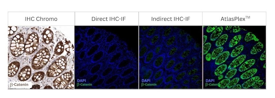
Figure 2. Comparison of β-catenin detection across four immunohistochemical staining methods. Human rectum tissue stained for β-catenin using the TripleA polyclonal anti-β-catenin antibody (Cat. HPA029159) and compared across four staining methods: classical DAB chromogenic IHC, direct IHC-IF, indirect IHC-IF, and AtlasPlex. The comparison demonstrates differences in staining intensity, specificity, and signal-to-noise ratio between the methods. AtlasPlex provides enhanced signal detection and improved contrast, enabling clear visualization of β-catenin expression in the tissue while maintaining the characteristic localization pattern.
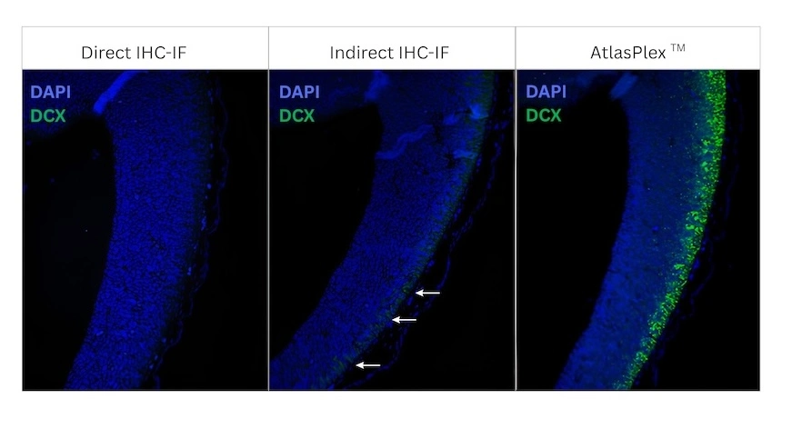
Figure 3. AtlasPlex enables sensitive detection of low-abundance DCX in the embryonic mouse cortex. Multiplexed immunohistochemical (IHC) staining of embryonic mouse cortex at embryonic day 11 (E11) using an anti-doublecortin (DCX) antibody (HPA036121). AtlasPlex signal amplification enhances the detection of the low-abundance neurodevelopmental marker DCX, resulting in a stronger and more distinct staining signal with improved signal-to-noise ratio. The amplified signal enables clear visualization of DCX-positive immature neurons in the developing cortex, demonstrating the utility of AtlasPlex for sensitive detection of low-expressed proteins in tissue sections.
6 Key Advantages of TSA in Multiplex IHC/IF
1. Enhanced Sensitivity for Low-Abundance Targets Because a single enzyme deposits an array of reporter molecules, TSA detects scarce proteins that direct or standard indirect detection methods miss entirely. This allows researchers to drastically reduce primary antibody concentrations while maintaining exceptional signal intensity.
2. Permanent, Non-Fading Covalent Bonds Because the tyramide reporter is covalently bound directly to the tissue matrix (rather than loosely held by an antibody-antigen interaction), the signal will not wash off during subsequent staining rounds. Slides retain high signal intensity for long-term archiving and re-imaging.
3. Crisp Spatial Resolution without Signal Diffusion Tyramide deposition is a tightly localized, rapid enzymatic reaction. The activated intermediates react so quickly that they cannot diffuse away from the enzyme site, preventing the "smeared halo" effect and delivering the sharp, high-contrast localization necessary for tumor microenvironment (TME) mapping.
4. Same-Species Antibody Multiplexing TSA removes the classic restriction against using multiple primary antibodies raised in the same host species (e.g., all mouse or all rabbit). Because the fluorophore is permanently bonded to the tissue, the HRP enzyme can be safely quenched between rounds, allowing the next same-species primary antibody to be applied without cross-reactivity.
5. Gentle Multiplexing Over Harsh Stripping Protocols Instead of using the aggressive, tissue-degrading stripping buffers required by older sequential protocols, TSA workflows utilize an iterative cycle: bind, amplify, and gently quench the enzyme. This protects delicate tissue morphology and preserves antigenicity across multi-marker panels.
6. High Signal-to-Noise Ratio on Standard Microscopes The combination of robust signal amplification and clean enzyme quenching keeps background autofluorescence exceptionally low. Furthermore, because the output is standard fluorescence, panels can be imaged on standard fluorescence microscopes without requiring specialized, high-cost multispectral imaging hardware.
Streamlining Spatial Biology: The AtlasPlex™ Multiplex Kit
While the benefits of TSA chemistry are undeniable, optimizing the protocol from scratch, including antibody titrations, conjugations, and quenching conditions, has historically been a major barrier to entry.
AtlasPlex eliminates the configuration bottleneck by packaging advanced TSA chemistry into a pre-validated, ready-to-use multiplexing kit:
- Rapid Workflows: Detect up to 5 markers on a single FFPE tissue section in one day (~1.5 hours per cycle).
- Fully Customizable Panels: Build custom 3-plex or 5-plex panels choosing from 12,000+ validated targets derived from the comprehensive Human Protein Atlas antibody portfolio.
- No Species Headaches: Eliminate secondary antibody steps entirely using pre-biotinylated primaries and same-species compatible TSA reagents.
- All-in-One Solution: Every box includes biotinylated antibodies, HRP labeling and quenching reagents, and premium tyramide dyes: no separate protocol assembly or sourcing required.
The true value of Tyramide Signal Amplification isn't just the chemistry. It is the biological context it reveals. AtlasPlex allows you to view more markers in true spatial context using the tissue and imaging equipment you already have, reproducibly, in a single day.