
You've got a highly validated antibody, a well-fixed section, and a clean protocol on paper. The stain still comes back weak, patchy, or drowning in background. Nine times out of ten, the antibody isn't the problem: the antigen retrieval step is.
What Is Antigen Retrieval?
Antigen retrieval is the process of restoring epitope accessibility by disrupting formalin-induced crosslinks through heat or enzymatic digestion.
When formaldehyde crosslinks alter the three-dimensional conformation of epitopes, even high-affinity antibodies cannot bind to their targets effectively. By deliberately breaking these crosslinks, antigen retrieval exposes the binding sites and enables accurate antibody recognition. Selecting the appropriate retrieval method is crucial for achieving consistent and reliable immunohistochemical results.
Formalin has been the standard fixative in histopathology since 1893, and it works by forming methylene crosslinks between proteins. Those crosslinks preserve tissue architecture beautifully, but they also fold epitopes into shapes your antibody no longer recognizes.
Antigen retrieval breaks those crosslinks back open. Skip it, get it wrong, or push it too far, and you'll see one of three outcomes: weak staining, no signal at all, or high background that reviewers will flag immediately.
Here are 9 tips for getting it right plus the checks we'd run before blaming the antibody.
1. Don't assume you need it
Antigen retrieval isn't universal. Alcohol-fixed frozen sections don't need it at all because alcohols don't mask epitopes the way crosslinking fixatives do. Fresh frozen sections generally only need a brief fix in ice-cold acetone (-20°C, 10–20 minutes). Retrieval is a formalin problem specifically; if your tissue was never crosslinked, don't add the step just out of habit.
2. Default to HIER, not PIER
Heat-Induced Epitope Retrieval (HIER) is the workhorse for FFPE tissue, and for good reason: Proteolytic-Induced Epitope Retrieval (PIER) is harder to control and more likely to damage morphology.
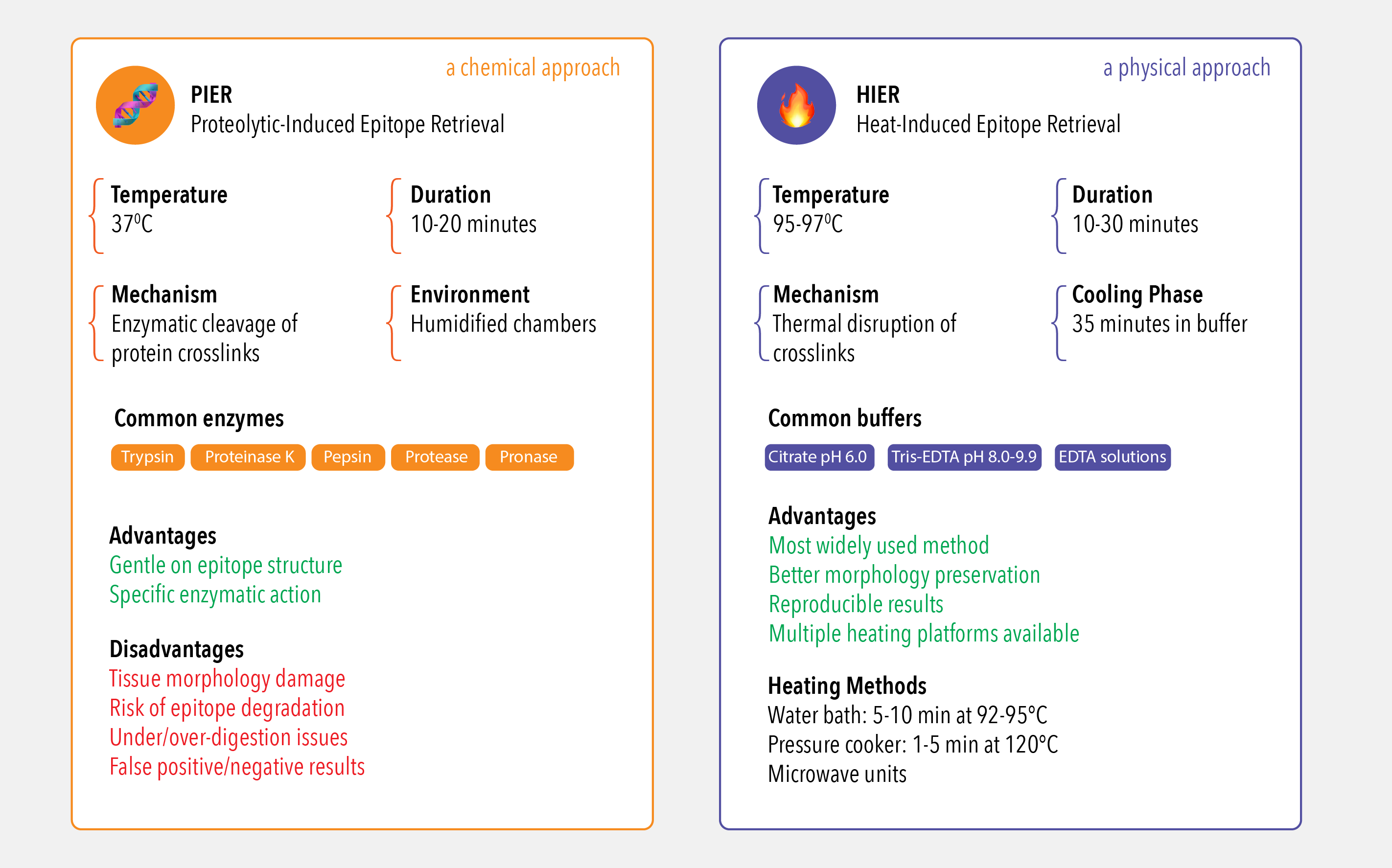
3. Test pH before you test time
pH matters more than most people optimize for. Start with both ends of the range in parallel — low pH citrate buffer (pH 6.0) and high pH Tris-EDTA (pH 8.0–9.9) rather than iterating on time first. It's faster to find your antigen's sweet spot by bracketing pH than by running the same buffer for progressively longer.
4. Match your heating platform to your throughput
Water bath, microwave, or pressure cooker all work, but the numbers aren't interchangeable: expect roughly 5–10 minutes at 92–95°C in a water bath versus 1–5 minutes at 120°C in a pressure cooker. Whichever you use, invest in temperature-controlled equipment: inconsistent heating is one of the most common causes of run-to-run variability, and it's invisible until you compare slides side by side.
5. If HIER genuinely fails, move to PIER
Some epitopes are heat-labile or sit in tissue that doesn't tolerate prolonged high heat well. That's when PIER earns its place, but treat it as a narrow tool, not a first resort. Digestion time is unforgiving: under-digest and you get weak, patchy staining; over-digest and you get false-positive background plus visible tissue damage. If you go this route, run a time-course in small increments rather than guessing a single endpoint.
6. Build controls into every run, not just validation runs
Three controls should be non-negotiable on every slide set:
Negative control: no primary antibody, to catch non-specific secondary binding
Positive control: tissue with known expression, to confirm the whole protocol is working
Specificity control: knockout/knockdown tissue or a blocking peptide, to confirm the antibody is binding the right target
7. Use a matched antigen as your gold-standard control
This is the control most labs don't know they have access to. If you're using an antibody where the manufacturer also sells the exact immunizing antigen spiking that antigen into your workflow confirms your antibody, your retrieval step, and your detection chemistry are all working together correctly. It's the closest thing to a positive control with zero ambiguity about what “working” means. We at Atlas Antibodies do this with our PrEST Antigens™ controls.
8. Diagnose problems by direction, not by guessing
When staining goes wrong, the fix depends on which way it failed:
Weak or no signal → under-retrieval. Increase heating time, or move to a higher-pH buffer before touching the antibody concentration.
High background → over-retrieval or tissue damage. Reduce time, drop to a lower pH, or check whether your secondary antibody is the actual source of noise.
Change one variable at a time. Changing pH and time together makes it impossible to know which fix actually worked.
9. Start from the datasheet, not from scratch
If the antibody you're using has a pre-optimized protocol from the manufacturer, use it as your starting point rather than reinventing conditions. At Atlas Antibodies, the large majority of our 12,000+ IHC-validated primary antibodies are optimized on a standardized HIER protocol specifically so you're not troubleshooting blind — check the datasheet before you build a matrix study from zero.
Atlas Antibodies
Your Partner in IHC Success
At Atlas Antibodies, we know that a high-quality antibody is only part of the equation for a successful IHC experiment. As an original antibody producer with roots in the Human Protein Atlas project, we've spent decades perfecting not just our antibodies, but the protocols that make them work. Antigen retrieval is one of the most critical steps, and getting it right is the key to unlocking clear, reproducible data. In this guide, we'll share our expertise on why it matters and how to perform it correctly.
Getting antigen retrieval right is what turns a validated antibody into a publishable result. If you're troubleshooting a specific target, our technical support team can help with protocol and buffer selection and if you want the closest thing to a guaranteed-working starting point, check whether your antibody has a matched PrEST Antigen™ available for a true positive control.