Cart 0


Immunohistochemistry (IHC)

Learn how our antibodies are validated and characterized in IHC and find the protocol you need to be successful in your IHC experiment.

Immunohistochemistry (IHC) is the most widely used application in histopathological diagnosis and research for the detection of proteins in tissues and cells. The use of antibodies to determine the presence and localization of specific proteins in surgical specimens is an indispensable tool in modern pathology.


Recommended Protocols 

In order to achieve the best results with our antibodies, make sure to use these recommended protocols. The protocols are optimized for our Triple A Polyclonals and PrecisA Monoclonals. Here you will also find our best advice for validation and troubleshooting in IHC.

IHC standard protocol
IHC protocol – Ventana Discovery XT


Validation and characterization in IHC

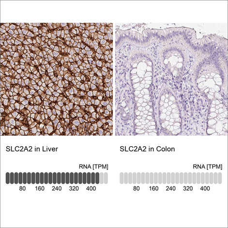
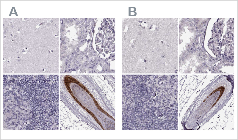
Our antibodies, Triple A Polyclonals, and PrecisA Monoclonals are characterized and validated by IHC-staining of formalin-fixed paraffin-embedded (FFPE) normal and cancer tissues in a tissue microarray format (TMA). 

Since IHC is a non-quantitative method our experienced personnel perform thorough manual analysis in an extensive number of human tissues to ensure specificity. 

The antibodies used for the complete profiling of protein expression in normal tissues from 144 different individuals corresponding to 44 normal tissue types and cancer tissues from 216 different patients (duplicate samples of tumor tissues representing the 20 most common forms of human cancer). In total, 576 tissue cores are immunostained and analyzed for each antibody. For the cancer tissues, an effort is made to include both high and low-grade malignancies. The images of the immunohistochemically stained tissues are manually annotated. Each tissue is assessed for staining intensity, localization, and fraction of stained cells. 


Anti-FKBP3 Antibody
Anti-FKBP3 Antibody

Anti-FKBP3 Antibody

HPA000864
In Stock (10+)
4 352 kr
Anti-JPH1 Antibody
Anti-JPH1 Antibody

Anti-JPH1 Antibody

HPA008996
In Stock (10+)
4 352 kr
Anti-SSB Antibody
Anti-SSB Antibody

Anti-SSB Antibody

HPA017287
In Stock (10+)
4 106 kr
Anti-FAM167A Antibody

Anti-FAM167A Antibody

HPA023915
In Stock (10+)
4 352 kr
Anti-ARHGAP18 Antibody

Anti-ARHGAP18 Antibody

HPA031595
In Stock (10+)
4 352 kr
Anti-TTC37 Antibody
Anti-TTC37 Antibody

Anti-TTC37 Antibody

HPA037905
In Stock (10+)
4 106 kr
Anti-DEFB107A Antibody

Anti-DEFB107A Antibody

HPA045626
In Stock (10+)
4 352 kr
Anti-CFAP100 Antibody

Anti-CFAP100 Antibody

HPA046354
In Stock (10+)
4 352 kr
Anti-SLC5A1 Antibody

Anti-SLC5A1 Antibody

HPA051805
In Stock (10+)
4 106 kr
Anti-KIAA0368 Antibody

Anti-KIAA0368 Antibody

HPA059795
In Stock (10+)
4 106 kr
Anti-FLT4 Antibody

Anti-FLT4 Antibody

HPA074389
In Stock (10+)
4 106 kr
Anti-CD3EAP Antibody

Anti-CD3EAP Antibody

HPA041664
In Stock (10+)
4 352 kr


Find the primary antibodies for your research

Triple A Polyclonals
PrecisA Monoclonals


Enhanced Validation in IHC

To further verify specificity, the validation performed for our antibodies is expanded with application-specific Enhanced Validation. In IHC, two different Enhanced Validation methods may be applied: 

  • Orthogonal validation (verification done with a non-antibody-based method
  • Independent antibody validation (two antibodies targeting the same protein but binding to different parts of the protein thus verifying each other)

Currently, over 5,000 antibodies are validated using at least one of these two enhanced validation methods. Learn more about the methods below. 


Orthogonal validation in IHC by comparing antibody signal to RNA

Orthogonal validation is an enhanced method for validation where the antibody staining is confirmed by a non-antibody-based method.

The method is applied in IHC by comparing the antibody staining to RNA-Seq data for the same samples, using both positive and negative samples. Antibody specificity is confirmed when the antibody signal in IHC matches the RNA levels.

For each antibody, two tissues are chosen for validation, one with high RNA expression and the other with low or no RNA expression of the target. The tissues are chosen so that there is at least a five-fold difference between the RNA expression in the high and low samples. 

In the validation data presented for the antibody, the IHC stainings from two samples are displayed together with their corresponding RNA values. 


Example of orthogonal validation in IHC. IHC staining of liver and kidney tissues using the Anti-SLC2A2 polyclonal antibody (HPA028997). The corresponding RNA-Seq data (TPM values) for the same tissues are presented below. Liver and colon samples were chosen because of their high and low SLC2A2 RNA expression, respectively.


Validation by independent antibodies in IHC

Validation by Independent Antibodies is an enhanced method for validation where the antibody specificity is demonstrated by comparing at least two antibodies targeting the same protein but with non-overlapping epitopes. 

If the staining patterns from the two antibodies correlate when compared across a panel of relevant tissues, the antibodies validate each other. At Atlas Antibodies, 20-44 tissues are evaluated for each antibody, and four representative tissues are displayed for each antibody. 

In the validation data presented for the antibody, the IHC stainings for both antibodies are displayed together. 


Example of validation by independent antibodies in IHC. The two Anti-TCHHL1 antibodies HPA063483 (left, A) and HPA042579 (right, B) target different regions of TCHHL1. The image shows IHC stainings of the two antibodies in the cerebral cortex, kidney, lymph node, and skin tissues. The antibody's staining pattern across positive and negative tissues is comparable, so the two antibodies validate each other.


All Protocols and guides

To achieve the best results with our antibodies, make sure to use these recommended protocols. The protocols are optimized for Triple A Polyclonals and PrecisA Monoclonals. See all our protocols for IHC, WB, and ICC-IF.

Go to protocols