The Challenge: Mapping the Brain's Cellular Complexity
The brain is one of the most cellularly diverse organs in the body, composed of specialized neurons and glial cells that coordinate everything from motor control to cognition. Understanding the cellular composition and spatial organization of the brain is essential for studying both normal function and disease mechanisms.
Multiplex immunohistochemistry (mIHC) enables detailed spatial profiling of multiple cell types within the same tissue section - an approach that is highly valuable for neuroscience research. However, traditional methods are often slow, complex, and limited by antibody cross-reactivity.
The AtlasPlex Solution: One-Day Spatial Profiling for Brain Tissue
Imagine being able to multiplex complex brain tissues without worrying about antibody host species, skipping secondary antibodies entirely, and completing the whole workflow in just one day.
This is exactly what AtlasPlex makes possible, Its optomized design, featuring biotinylated primary antibodies and powerful tyramide signal amplification (TSA), delivers clear, robust results for high-impact neuroscience research.
Let’s explore two powerful applications:
Case Study 1: Profiling Neuronal and Glial Cells in the Adult Human Cerebellum
The ability to visualize neurons, astrocytes, oligodendrocytes, and microglia simultaneously provides valuable insight into how different cell types interact in both health and disease. For example, alterations in astrocyte morphology or microglial activation can reflect early neuroinflammatory changes, while loss of Purkinje neurons contributes directly to motor dysfunction in cerebellar degenerative diseases.
In this example, AtlasPlex was used to perform a single-round 5-plex mIHC experiment on FFPE human cerebellum, enabling simultaneous visualization of neuronal and glial cell populations without secondary antibodies or antibody species constraints—all within a one-day workflow.
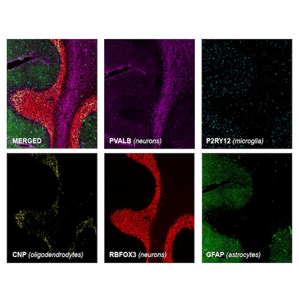
Cerebellum 5-Plex Panel Configuration
| Target Cell Type | Marker | Product Number | Biological Significance |
|---|---|---|---|
| Neurons | RBFOX3/NeuN | HPA030790 | Definitive marker for mature neurons |
| Neurons | PVALB | HPA048536 | Identifies calcium-binding interneurons |
| Astrocytes | GFAP | HPA056030 | Key marker for macroglial cells & injury response |
| Oligodendrocytes | CNP | HPA023280 | Highlights myelin-producing cells |
| Microglia | P2RY12 | HPA014518 | Identifies homeostatic microglial states |
This mIHC panel illustrates how AtlasPlex can resolve the organization of neuronal and glial populations within the human cerebellum, a region critical for motor coordination, learning, and cognitive processing. Disruptions in cerebellar cellular architecture have been implicated in a range of neurological conditions, including spinocerebellar ataxias, multiple system atrophy (MSA), and autism spectrum disorders.
Blog: AtlasPlex Multiplex IHC: From FFPE Sample to 3-Plex Data in a Single Day
Case Study 2: Mapping Neurogenesis in the Developing Brain
Brain development is a complex process, as includes neuro- and gliogenesis, neuronal cell maturation and migration, and establishing synaptic connections. Cell-type specific markers are a useful tool for studying nervous system development in both health and disease.
During development, the mouse brain undergoes rapid changes in cellular composition and spatial organization. AtlasPlex enables the simultaneous detection of neuroepithelial cells and neuronal subpopulations within the same tissue section, providing a high-resolution view of neurogenesis and early cortical patterning. This approach is crucial for studying developmental brain disorders such as microcephaly, lissencephaly, and cerebellar hypoplasia, which are linked to disruptions in neuronal differentiation and migration.
👇🏼 In this example, AtlasPlex was used to perform a single-round 3-plex mIHC experiment on E14 mouse embryo brain tissue, enabling simultaneous visualization of neuroepithelial cells, immature neurons, and early-born cortical neurons without secondary antibodies or antibody species constraints. Using markers for SOX2, DCX, and TBR1, this panel provides a spatial snapshot of early cortical development, capturing progenitor populations alongside differentiating and migrating neurons within the same tissue section in a one-day workflow.
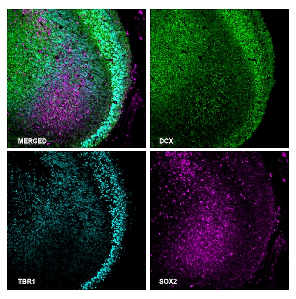
Neurogenesis 3-Plex Panel Configuration:
- Neuroepithelial Cells: Anti-SOX2 antibody (Cat. AMAb91307)
- Immature Neurons: Anti-DCX antibody (Cat. HPA036121)
- Early-born Cortical Neurons: Anti-TBR1 antibody (Cat. HPA078644)
By simultaneously detecting SOX2, DCX, and TBR1, AtlasPlex provided a high-resolution snapshot of early cortical development. Researchers can see progenitor populations alongside differentiating neurons as they migrate to form the cortical plate, eliminating the "alignment error" inherent in serial sectioning.
Blog: AtlasPlex: Design Multiplex IHC Panels with an Unrivaled Choice of 12,000 HPA Targets
🧠 Advancing Neuroscience Through Multiplex Imaging
AtlasPlex mIHC technology offers a scalable and reproducible way to explore brain tissue complexity across developmental stages and disease models. In neuroscience, where cellular heterogeneity and spatial context are critical, the ability to simultaneously visualize multiple markers within the same tissue section provides new opportunities to link molecular pathways with histological phenotypes.
Whether applied to human or rodents brain, AtlasPlex facilitates deeper understanding of cellular networks and their alterations in neurological disease. As research into neurodegenerative, neurodevelopmental, and neuroinflammatory disorders accelerates, multiplex imaging platforms like AtlasPlex are becoming essential tools for translating molecular data into biological insight.
Illustrative examples of neuroscience-focused AtlasPlex panels include:
- Synaptic and neuronal activity panels (e.g. SYN1, SLC17A7, GAD1),
- Myelination and white-matter integrity panels (e.g. MBP, PLP1, OLIG2),
- Blood–brain barrier or neurovascular unit panels (e.g. CLDN5, PECAM1, PDGFRB)
- Neuronal subtype specification panels (e.g. SATB2, TBR2/EOMES, FOXP2)
- Astrocyte functional state panels (e.g. ALDH1L1, SLC1A3/GLAST, S100B)
- Neuroinflammation and microglial activation panels (e.g. CX3CR1, TMEM119, CD68)
Importantly, these are only illustrative examples: AtlasPlex panels can be fully customized by choosing from over 12,000 validated protein targets, giving you the freedom to design marker combinations tailored to your specific brain region, disease model, or biological question.
Why Choose AtlasPlex for Your Neuroscience Research?
The AtlasPlex kit brings together several smart features to simplify and accelerate multiplex immunohistochemistry (mIHC) workflows with several key advantages:
- Total Panel Freedom: Build fully custom 3-plex or 5-plex panels from a catolog of over 12,000 validated antibodies published on the Human Protein Atlas, eliminating antibody host-species restrictions.
- Enhanced Signal Strength: the workflow combines biotinylated primary antibodies with tyramide signal amplification (TSA) for bright, stable signals even for low-abundance proteins.
- No secondary antibodies: By removing the need for secondary antibodies, you simplify panel design and minimize the risk of cross-reactivity.
- Fast, Gentle Workflow: Replace harsh, multi-day stripping protocols with a gentle HRP quenching process that preserves tissue morphology. Go from sample to data in a single day
Frequently Asked Questions (FAQ)
Q1: What is AtlasPlex? AtlasPlex is a complete multiplex immunohistochemistry (mIHC) kit designed for the rapid and customizable spatial profiling of 3 to 5 protein targets in a single tissue section.
Q2: How does AtlasPlex prevent antibody cross-reactivity? The workflow uses biotinylated primary antibodies and a gentle HRP quenching step after each staining cycle. This approach eliminates the need for secondary antibodies and harsh antibody stripping, which are common sources of cross-reactivity and tissue damage.
Q3: Is AtlasPlex compatible with both human and mouse tissue? Yes, AtlasPlex is highly effective for both human and rodent brain tissue, as demonstrated in the case studies. You can select from thousands of validated antibodies specific to your species of interest.
Blog: Multiplexing Q&A: From Complexity to Clarity with AtlasPlex
Explore AtlasPlex NeuroScience Solutions